
Psychosis is a mental state in which a person loses touch with reality. During a psychotic episode, perception and thinking can become disrupted, making it difficult to distinguish what is real from what is not. Common signs of psychosis include disorganized speech or thought patterns, delusions—false beliefs often rooted in fear or suspicion—hallucinations, which involve hearing, seeing, or sensing things that aren’t there, and thoughts that jump rapidly between unrelated topics.
Studies suggest that the prevalence of psychosis is between 15 and 100 people per 100,000 people. It most often emerges in late adolescence or early adulthood, typically between the late teens and mid-20s, though it can occur at younger or older ages.
While psychosis is most commonly associated with schizophrenia, it can also occur in some individuals with bipolar disorder, severe depression, or certain personality disorders. In addition, psychotic episodes may be triggered by medical or external factors. These can include alcohol or drug use and withdrawal, brain disorders, brain tumours or cysts, and certain medications.
Psychosis was previously thought to be a genetic risk, however it is now considered a heterogenous spectrum with multiple underlying causes and manifestations[1].
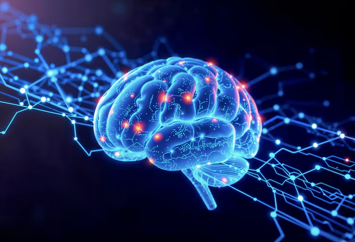
Some studies have found that psychosis is due to an imbalance of dopamine in the brain whereas others focus on the cortical brain and its structure rather than chemical imbalances.
Another 10 year longitudinal study measured cortical thickness as a biomarker to track the clinical trajectories over time[2].
The neurodegenerative explanation for schizophrenia proposes that psychosis results from the progressive, ongoing deterioration of the brain (similar to Alzheimer’s) where gray matter loss and cortical thinning steadily worsen over time and therefore lead to increasingly severe symptoms. This study challenges this view by showing that cortical abnormalities in individuals with schizophrenia did not follow a consistent downward trajectory; instead, they appeared irregular and the symptoms reduced over time. This pattern contradicts the idea of continuous neurodegeneration and instead suggests that schizophrenia may not be primarily a degenerative disease, opening the door to alternative interpretations such as neurodevelopmental or other perspectives.
Another study on the abnormalities in the cortical brain found that there are brain cortical alterations in individuals with psychotic disorders. Compared to healthy individuals, individuals with schizophrenia show widespread thinning of the cortex and a reduction in cortical surface area, with the most pronounced differences in the frontal and temporal lobes. Even when overall cortical thickness is taken into account, these regional differences remain significant, suggesting that some areas of the brain are particularly affected, while reductions in surface area tend to be more global. Cortical thinning was notably greater (around two to three times larger) in patients taking antipsychotic medications compared with those who were unmedicated. In addition, age-related thinning was more pronounced in individuals with schizophrenia than in healthy controls. Cortical thickness in specific regions was also linked to other clinical factors: it decreased with higher medication doses, more severe symptoms, and longer illness duration, but increased with later age at onset [3].
This is further supported through a study showcasing that individuals at clinical high risk who have increased psychotic symptoms have been found to have a faster rate of cortical grey matter decrease than those without symptomatic progression or healthy control subjects. However, it is unclear whether such alterations reflect mechanisms linked with schizophrenia’s aetiology or antipsychotic medication exposure [4].
While psychosis is often diagnosed through clinical assessment alone, lab tests and brain scans can provide valuable additional information. Treatment typically involves antipsychotic medication, cognitive behavioural therapy, or other forms of psychotherapy, depending on the individual’s needs. Despite advances in understanding and managing psychosis, much remains to be learned about its underlying mechanisms and complexities, highlighting the need for ongoing research.
References:
Scott, J. G. (2025). Annual Research Review: Psychosis in children and adolescents – a call to action: a commentary on Kelleher (2025). Journal of Child Psychology and Psychiatry, 66(4), 588–591.
Berthet, P., Haatveit, B. C., Rikka Kjelkenes, Worker, A., Kia, S. M., Wolfers, T., Rutherford, S., Dag Alnaes, Dinga, R., Pedersen, M. L., Dahl, A., Fernandez-Cabello, S., Dazzan, P., Agartz, I., Ragnar Nesvåg, Ueland, T., Andreassen, O. A., Simonsen, C., Westlye, L. T., & Melle, I. (2024). A 10-Year Longitudinal Study of Brain Cortical Thickness in People with First-Episode Psychosis Using Normative Models. Schizophrenia Bulletin, 51(1), 95–107.
van Erp, T. G. M., Walton, E., Hibar, D. P., Schmaal, L., Jiang, W., Glahn, D. C., Pearlson, G. D., Yao, N., Fukunaga, M., Hashimoto, R., Okada, N., Yamamori, H., Bustillo, J. R., Clark, V. P., Agartz, I., Mueller, B. A., Cahn, W., de Zwarte, S. M. C., Hulshoff Pol, H. E., & Kahn, R. S. (2018). Cortical Brain Abnormalities in 4474 Individuals With Schizophrenia and 5098 Control Subjects via the Enhancing Neuro Imaging Genetics Through
Cannon, T. D., Chung, Y., He, G., Sun, D., Jacobson, A., van Erp, T. G. M., McEwen, S., Addington, J., Bearden, C. E., Cadenhead, K., Cornblatt, B., Mathalon, D. H., McGlashan, T., Perkins, D., Jeffries, C., Seidman, L. J., Tsuang, M., Walker, E., Woods, S. W., & Heinssen, R. (2015). Progressive Reduction in Cortical Thickness as Psychosis Develops: A Multisite Longitudinal Neuroimaging Study of Youth at Elevated Clinical Risk. Biological Psychiatry, 77(2), 147–157.